atanatomy.weebly.com

Book II: Child Development

Home

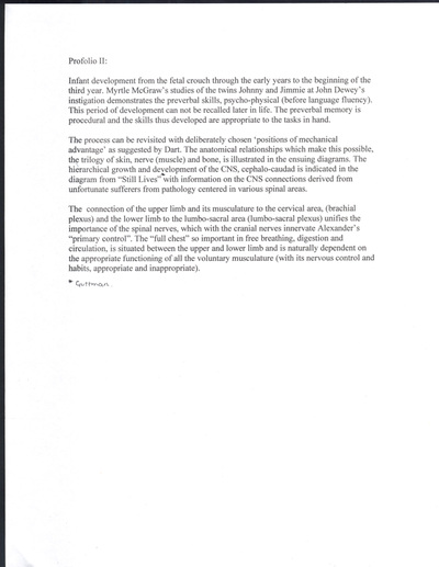
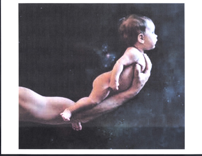
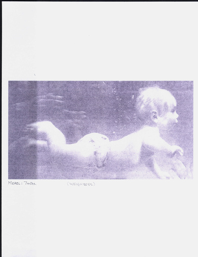
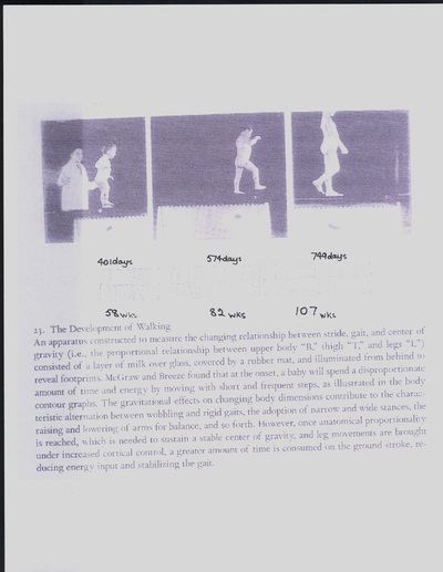
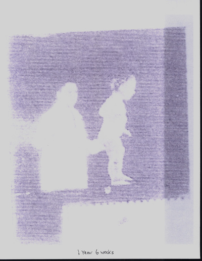
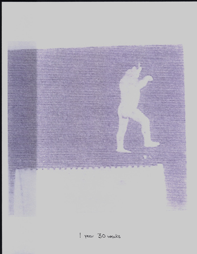
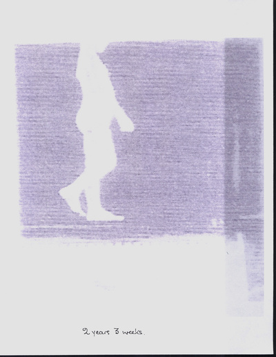
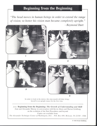
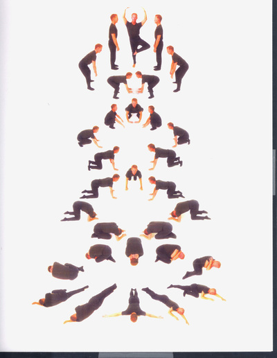
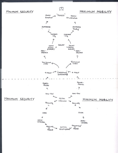
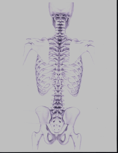
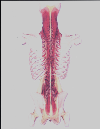
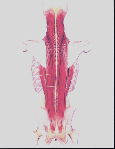
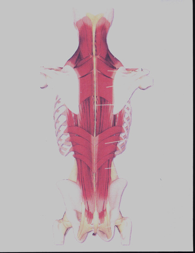
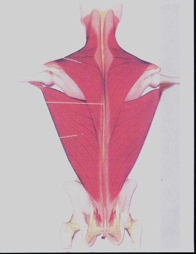
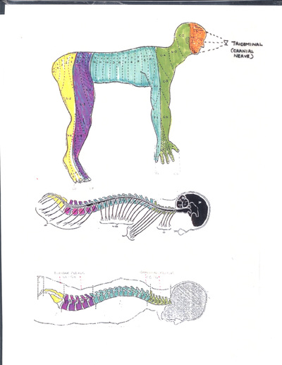
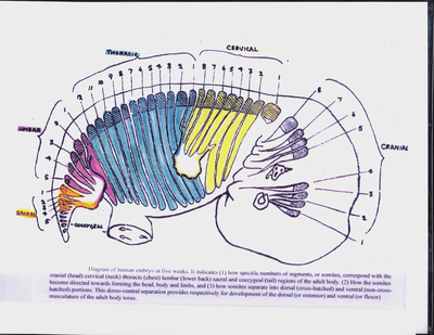
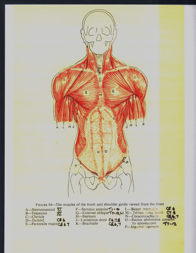
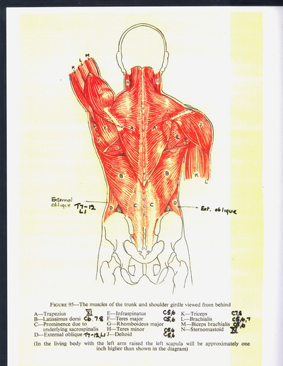
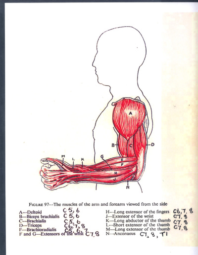
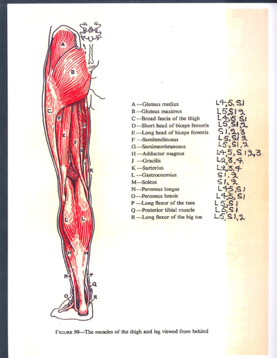
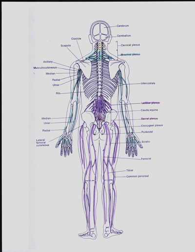
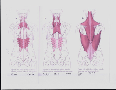
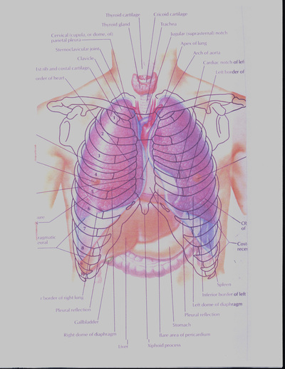
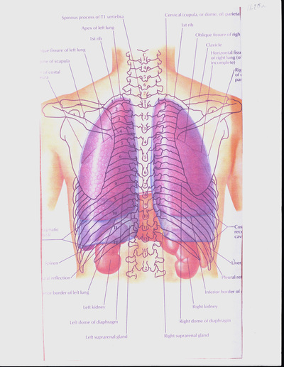
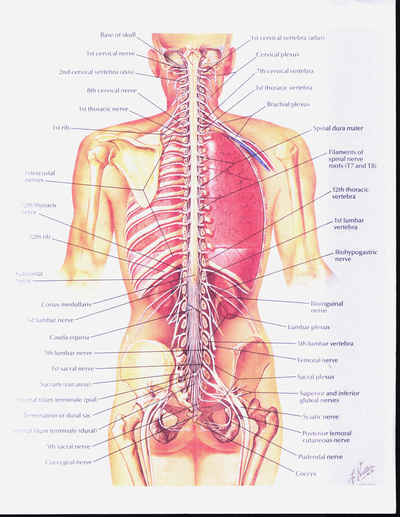
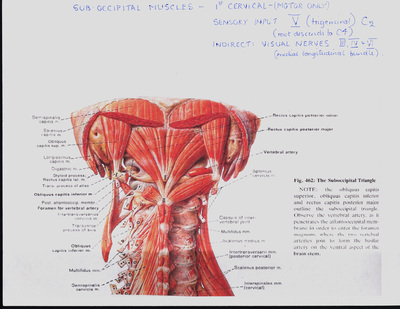
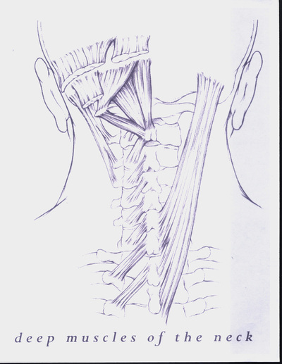
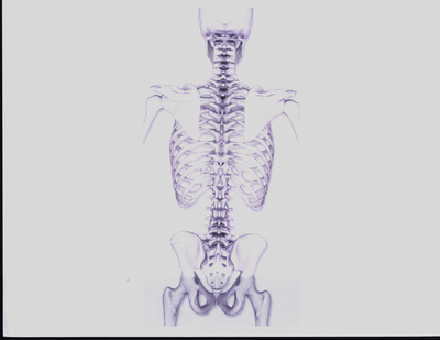
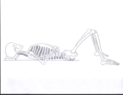
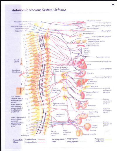
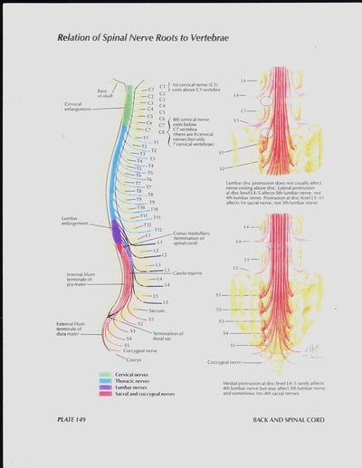
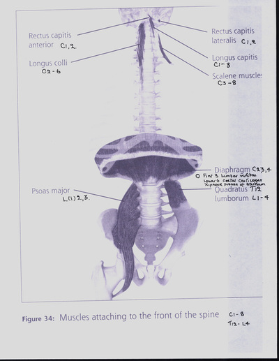
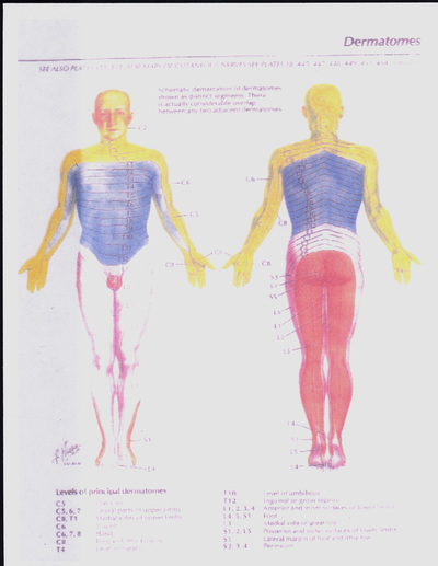
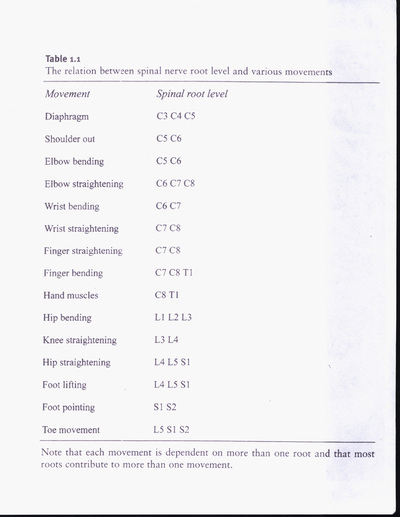
Book II presents child development, from birth to age 3, relying heavily on Myrtle McGraw's studies of Johnny and Jimmy. This process, which Raymond Dart argues is usually lost in the adult's “bespectacled decrepitude,” is revisited with the Alexander Technique. Also presented is the neuro-muscular-skeletal system, particularly the spinal and cranial nerves, as they are the underpinnings of this developmental process, and attempts later in life to relearn it.

View Book II as Slide Show

II.1 Introductory notes (Murray)
II.2 Fetal thumb sucking
II.3 Fetal position (infant)
II.4 Fetal position (adult)
II.5 Fetal position (adult)
II.6 Infant, extension, 3.5 months
II.7 Infant, extension, aided
II.8 Infant, underwater
II.9 Birth spiral
II.10 Developmental Profile, from Institute for the Advancement of Human Potential (IAHP)
II.11 Infant walking, aided
II.12 Infant walking, aided by F.M. Alexander
II.13 Child crawling, standing, walking
II.14 Walking development, 11-37 weeks (McGraw)
II.15 Walking development, 58-107 weeks (McGraw)
II.16 Walking development, 58 weeks (McGraw)
II.17 Walking development, 82 weeks (McGraw)
II.18 Walking development, 107 weeks (McGraw)
II.19 Prone locomotion development
II.20 IAHP Sensory Development, from IAHP Developmental Profile
II.21 Johnny, on boxes (McGraw)
II.22 Johnny on steep slide (McGraw)
II.23 Walking development, 11-107 weeks (McGraw)
II.24 Baby looking, (Goldberg, Ahern, Murray)
II.25 Vertebrate pattern of evolution (Halstead)
II.26 Evolution and The Attainment of Poise (Dart)
II.27 Dart-Alexander Procedure (Murray)
II.28 Dart Procedure, fetal upwards (Johnston)
II.29 Dart Procedure: fetal upwards, mobility vs security
II.30 Skeleton of head, neck, and back: posterior view
II.31 Back musculature, interspinal (layer 1, deepest layer)
II.32 Back musculature, longissimus (layer 2)
II.33 Back musculature (layers 3 and 4)
II.34 Back musculature, trapezius and latissimus (layer 5)
II.35 Dermatomes (quadrupedal), with CNS and skeleton (bone, nerve, skin)
II.36 Embryo segmentation, 5 weeks (Dart)
II.37 Superficial muscles of trunk and shoulder girdle: front view (RAF)
II.38 Superficial muscles of trunk and shoulder girdle: back view (RAF)
II.39 Arm and forearm muscles: side view (RAF)
II.40 Dermatomes, upper limb (Netter)
II.41 Dermatomes and movements of lower limbs (Netter)
II.42 Thigh and leg muscles: side view (RAF)
II.43 Thigh and leg muscles: front view (RAF)
II.44 Thigh and leg muscles: back view (RAF)
II.45 Central nervous system: spinal nerves plexuses
II.46 Back musculature: layers 3, 4, and 5 (Dimon)
II.47 Lungs: front view
II.48 Lungs: back view
II.49 CNS and nerve plexuses (Netter)
II.50 Suboccipital muscles (Netter)
II.51 Muscles of the neck (superficial)
II.52 Muscles of the neck (deep)
II.53 Neck, external features in relation to underlying structures
II.54 Suboccipital muscles
II.55 Atlas and axis (Dimon)
II.56 Cervical spinal column
II.57 Muscles attaching front of spine
II.58 Hip and torso muscular system
II.59 Spinal movement, pulley mechanism (Grundy)
II.60 Muscles attaching front of spine (Dimon)
II.61 Skeleton of head, neck, and back: posterior view
II.62 Skeleton: semi-supine
II.63 Spinal curves
II.64 Hip and knee joint structure
II.65 Joints
II.66 Autonomic nervous system
II.67 Nerves: spinal nerve roots and relation to vertebrae (Netter)
II.68 Types of spinal movement
II.69 Back: F.M. Alexander (Man's Supreme Inheritance)
II.70 Release of psoas
II.71 Release of psoas
II.72 Leg musculature
II.73 Torso and leg musculature
II.74 Hip musculature: external rotators
II.75 Abdominal wall: posterior view (Netter)
II.76 Lumbar nerve plexus (Netter)
II.77 Sacral and coccygeal nerve plexuses (Netter)
II.78 Muscles attaching front of spine (Dimon)
II.79 Frog saltation
II.80 Amphibian skeletal evolution
II.81 Dermatomes, full body, anterior and posterior (Netter)
II.82 Femur angles
II.83 Pelvis, angles and weight bearing
II.84 Hip: seated (Dimon)
II.85 Knees forward and away, excerpt (Rugg-Gunn)
II.86 Relation between spinal nerve root and movement (Cole)
Powered by Create your own unique website with customizable templates.